


Onychomycosis(nail fungus) is an damage to the fungal infection of the nail plate and the structures around it: nail rollers, matrix (nail seed) and nail bed.It is manifested by the deformation and thickening of the nails, a change in their colors - the nails become white or yellow.
This disease is often found.The prevalence of onycomycosis in Europeans, according to some reports, reaches 10-12 %, exceeding the well-known indicators of the last decade.In men, it happens 1.5 times more often, but they contact the doctor 2 times less frequently than women.Older people get sick more often, children are very rare.
The main problem in treating the disease is that patients come to see a dermatologist for a long time after the appearance of the first symptoms.Because of this, pathological fungi capture a large area and treatment is delayed.
Pathogenic fungi can only be transmitted by a sick person.Very often, infection with a fungus occurs within the family, as the source is not detected in time and adequate preventive measures are taken.
Causes of the disease:Most often, direct contact with the patient or objects he uses (shoes, clothes, baths, washing clothes, manicure supplies).Often the infection occurs when you visit gyms, baths, saunas and pools.
The development of the disease is facilitated by microscience - cracks in interdigital folds that occur due to boredom, increased sweating, dry skin, poor drying after water procedures and flat legs.
Nail mycoses can also occur in the presence of concomitant diseases of the endocrine system (diabetes, overweight, hypothyroidism), vascular diseases of the limbs (venous deficiency, lymphostases), immune disorders, as well as using antibiotics, cytostatic medicines and cytostatic.As a result of the above diseases, the blood microcyloration in the nail area is disturbed and the natural immunity is reduced, which contributes to the development of secondary infection.
Onychomycosis cause the following types of fungi:
- dermatophyte;
- Candida peak -like mushrooms;
- Casting.
Depending on the type of pathogen, the penetration of a fungus infection and the clinical appearance are different, so approaches to therapy are also different.
Foot nails are affected by a fungus 10 times more often than in the hand.In most cases, dermatophytes cause fungi (for example, Trichophyton rubrum).The remaining cases are often caused by contradictory molds (aspergillus, scopulariopsis, fusarium).
If similar symptoms are detected, consult a doctor.Don't have it yourself -My - it's dangerous to your health!
Symptoms of onycomycosis (nail fungus)
The longer the disease persists, the more pronounced its symptoms become.The main signs of onychomycosis include:
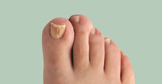
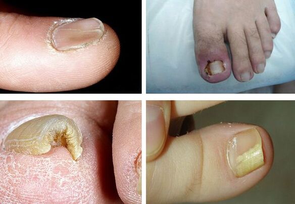
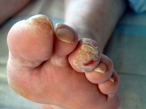
- Dischromia - a change in the color of the nail in yellow, black, green, gray or brown (the type of color depends on the type of fungus);
- Onikoliza - separation of nail plate from the box;
- the change in the thickness of the nail plate;
- Gapalonichia - a power of tile thickness and its softening;
- Koulonichia - the nail looks concave, in the form of a teaspoon;
- Pahionichia - nail plate obesity, nail hypertrophy;
- Onichogrifosis - obesity, a change in the color of the nail plate, curved in the shape of a beak;
- change in nail bed thickness (hyperkeratosis - nail bed thickening);
- Changing the surface of the nail plate: pits, furrows, ridges;
- Changing the nail rotators and surrounding skin (paronychy - inflammation of the proximal nail roller).
It is important to note that none of the symptoms is pathogen, that is, vaguely appropriate for a particular pathogen, so it is impossible to determine with symptoms - additional examinations are required.
Pathogenesis of onycomycosis (nail fungus)
The pathogenesis of the disease depends on how the fungus strikes the skin and nails.
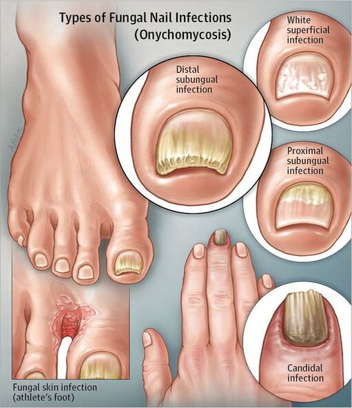
Distal type of submarine:If the fungus is inserted through the skin in the area of the nail or distal region, then the spread of the infection occurs through the free edge of the nail in the bed and further on the matrix.At first, the nail plate may not change, but later, due to hyperkeratosis, gradually removing from the nail bed and becoming yellow.Gradually, a thickening of the nail plate is possible.
White Surface Type:If white focus forms on the surface of the nail, then over time, the fungus of the entire nail plate occurs.The nail thickens, calls, acquires a gray-brown tinge.In this case, the nail bed matrix and epithelium are not affected.There is no inflammation of the surrounding skin.
Proximal type of under -fire:The fungus can be spread from the skin and periological rollers on the nail plate and then to the matrix, reaching the distal parts of the nail plate.Spots appear on the nail in the area of the hole and the nail bed, the nail plate is disconnected.There is no pronounced inflammation of the nail bed or matrix.
Total Distrophic Type:The whole nail is touched.The proximal areas of the nail roll disappear or thicken, so the nail plate can no longer form and grow.
thereThe biophysical concept of the pathogenesis of onycomycosis, which says that with the disease, the confrontation between the two forces occurs: a fungus colony that grows in the direction of the matrix and the natural growth of the nail from the matrix to the distal edge.Therefore, the rate of nail growth is essential during onycomycosis - the sooner the nail grows, the earlier the treatment comes.Perhaps this is exactly what explains the small spread of the disease in children, as their nails grow faster than in adults and older people.
Classification and stages of onycomycosis development (nail fungus)
There is the following classification of onychomycosis:
- Distal submarine;
- superficial white;
- Under -proximal gear;
- Total dystrophic.
According to the 1970 classification:
- Normal: In the nail thickness, a yellow and white ribbon, but the shape of the nail plate does not change, there is no submarine hyperkeratosis;
- Hypertrophic:The nail plate turns yellow, thickens due to submarine hyperkeratosis, becomes broken, with clumsy edges;
- Distrophic:There is a thinning and removal of the nail plate from the nail bed with the formation of gaps.
Complications of onycomycosis (nail fungus)
With long existence onycomycosis, the risk of development increasesdiabetic(forming trophic ulcers standing) andgangrene, if the patient has diabetes or vascular disease of the lower extremities.
In immunosuppressive conditions (primary and secondary immunodefof), fungi can spread to the skin, internal organs and cause body allergies.This can be manifested by rashes on the skin until the development of bronchial asthma.
Diagnosis of onycomycosis (nail fungus)
Before dismantling the methods of diagnosing onycomycosis, it is necessary to explain how to properly accumulate the material for the study (the patient makes it independently, or prepares the nails before diagnosis).Before the fence of the material for the study, it is necessary to treat the nail plate with 70 % alcohol so that there is no obstruction of other bacteria.
The method of collecting the material varies depending on the form of onychomycosis:
- Superficial form- make a piece from the nail plate;
- Distant form- a piece of nail bed is needed and a piece of nail plate is needed;
- Proximal submarine form- The material is harvested by a drill, or a nail biopsy, or a piece of nail bed, is made.
The fastest method for determining the pathological fungi on the nail ismicroscopy.Technique: The studied material is treated with an alkali solution for the distribution of keratin.To make the strands of the best mushrooms, the paint is added to the alkali.Next, study the resulting medicine under a microscope.
This method of research is faster and more objective.Sensitivity is up to 80 %.The disadvantages of the method include the fact that when using it, it is impossible to determine the type of pathogen.
Bacteriological planting: It is an additional method of diagnosing onycomycosis.The material is planted in a separate environment and the result is interpreted under a microscope after 2-3 weeks.This method allows you to create the type of pathogen - this helps to determine the treatment tactics and when choosing medication by sensitivity.But the disadvantage of the study is that it takes a long time, and its sensitivity is only 30-50 %.
Biopsy: With the help of a scalp and with the use of anesthesia, a nail and a nail bed are cut.The material is immersed in a formaldehyde solution and sent for histological examination in the laboratory.The advantages of this method are very sensitive and allow you to determine the presence of a pathological fungus in the material.
Minor Offenses: It is impossible to identify the pathogen, as well as to determine the sustainability of microorganisms, the high cost and complexity of the method.
Genodiagnostics: Molecular Method of Biological Research (PCR).This is one of the new and very sensitive methods for the diagnosis of onychomycosis-with its help, the DNA of the pathogen is detected.It is recommended to present this type of diagnosis in those medical institutions that have PCR laboratories, but at the moment, test systems to identify dermatophytes and mold fungi only plan to present to the laboratory.The method allows you to determine the type of pathogen, and its sensitivity is from 80-90 %.Cons - high cost, inaccessibility, lack of technology standards and complexity of execution.
More and more doctors are presented in practicedermatoscopy.Using this method, you can evaluate a change in the color and structure of the nail, the state of the surrounding structures.Dermatoscopic examination allows you to more accurately evaluate the depth of nail plate damage and more accurately calculate the severity of onycomycosis.
Treatment of onycomycosis (nail fungus)
There are several types of treatment for onycomycosis:
- Local therapy.
- System therapy.
- Combined therapy.
- Corrective therapy.
Local therapyIt involves applying medication to the nail plate and nail rollers.Indications for local therapy:
- Limited form of nail plate damage.
- There are contraindications for the appointment of systemic medicines: hypersensitivity, liver disease, kidney function, pregnancy, lactation.
The advantages of this therapy are that high concentrations of a therapeutic agent form on the surface of the nail, which does not penetrate the bloodstream.There are no side effects of using antifungal drugs - nausea, a decrease in appetite, abdominal pain.The disadvantage of the method is that the medicinal substance does not always fall into the pathogen's habitat, especially if the fungus is located on the nail bed or the matrix.This, in turn, can lead to inefficiency of treatment.This type of treatment is a long time, as before applying the medicine, it is necessary to remove the affected part of the nail.
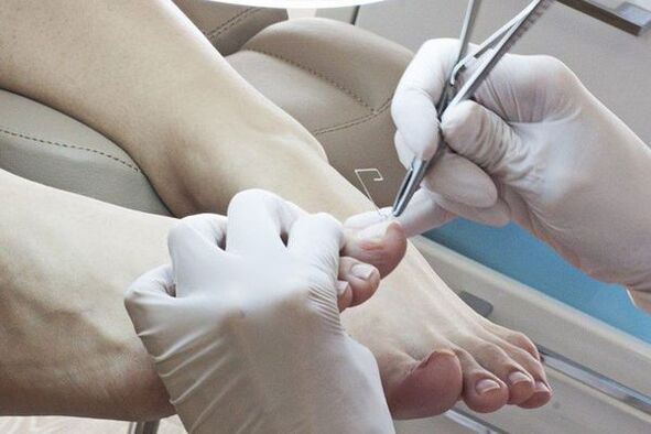
Ways to remove affected nails:
- Mechanical removal with files, nippers or drills.
- With the help of keratolytic plaster.Before applying a piece of keratolytic, the skin around the nail is closed with a patch, a patch (urea with salicylic acid) is applied to the top and sealed with an adhesive plaster.The patch mass is changed every 2-3 days.After each removal, the affected part of the nail is mechanically removed.
- Surgical this surgery is very painful and traumatic, as when removing the nail plate, the seedling area can be damaged, which leads to the growth of deformed nails.
Local antifungal agents are used after removing the affected nail plate.antimycoticDistinguish between the place of application:
- Applied to the nail: curves;
- Apply for rollers: creams, ointments, solutions.
The most studied tool for local use is a 1 % solution of the alllamin group drug, which has a test basis for treatment based on the meta-analysis of Cocranny.This tool has a water base, which contributes to the better penetration of the antifungal substance at the site of destruction.Lacs have a dehydrated base that reduces the penetration of the drug into deep layers.Therefore, dermatologists consider the use of unsatisfactory spray and increasingly prefer a water -based tool.
To get the result from local therapy, it is necessary to observe the treatment regimen, it is important for the patient to be responsible, stable and patient.The duration of therapy can reach 12 months.
System therapyIt allows the antifungal drug to penetrate the blood into the lesions, even if the nail bed and matrix are affected.The high concentration of the drug remains for a long time in lesions after the end of use.The disadvantages of this type of treatment are associated with the risk of side and toxic effects.
Indications for systemic therapy:
- Common forms of nail plate damage.
- Lack of effect of local therapy (that is, after six months of treatment of onycomycosis in the hands and 9-12 months of treatment of onycomycosis of the feet, had no healthy nails growing).
To determine treatment tactics, a clinical index of the severity of onycomycosis is used.It is used as a therapeutic standard in different parts of the world.
medicamentsFor the treatment of onychomycosis, you can classify as follows:
- Antimycotics - has an antifungal effect;
- Antiseptics - have antifungal and antibacterial effects.They are rarely used only if there are no other antifungal agents;
- Multicomponent - in addition to an antifungal agent, contain other medicines, such as anti -inflammatory.
Drugs to prescribe medicines:
- Standard - daily administration of drugs during the specified treatment period;
- Shortened - the treatment period is shortened, can be performed with conventional doses or enlarged;
- Intermittent - treatment is described in some short courses, intervals between courses are equal to the duration of the courses;
- Pulse therapy-Treatment is described in some short courses, intervals between courses are larger than the duration of the courses.
Antifungal drug is divided into active substance:
- Triazols;
- Allamine;
- Morpholines.
Currently, system therapy is usedOnly third generation drugs.
With combined therapyLocal and systemic treatment is performed simultaneously.Combined therapy is used if it is necessary to increase the effectiveness of systemic therapy and reduce treatment periods.
Corrective therapy(Treatment of accompanying diseases): In order to choose a treatment regimen, it is necessary to evaluate the general somatic condition of the body.Diseases such as circulatory disorders in the limbs can reduce the entry of the antifungal agent into the lesion.Therefore, medicines that improve trophic tissue are prescribed.
Due to the toxic effects of systemic antifungal drugs, it is necessary to exclude liver diseases and, if necessary, to prescribe hepatoprotectors.
Forecast.PREVENTION
The sooner the patient contacts the doctor with signs of fungal nail lesions, the sooner the disease will heal and restore the nail plate.With existing long processes with the capture of the entire nail, the treatment of onycomycosis can be long, but according to all recommendations, recovery often occurs.If there are contraindications to systemic therapy, long -term support treatment with local medicines is needed.
For preventionIt is necessary to comply with the rules of personal hygiene and reduce the new chance of infection:
- Try to wear comfortable and high quality shoes (in order to prevent increased foot sweating);
- It is recommended to change socks and tights daily;
- Use only individual shoes.Those undergoing treatment for onycomycosis should be treated at the beginning of treatment, at least once a month throughout the treatment period and after its completion;
- If necessary, use antiperspirants for the feet;
- Use an individual set of nail care (scissors, saws);
- Before and after visiting public places (pool, bathroom, sports hall), use external antifungal products (spray, creams and pencils);
- To identify the source of fungal infection in the family and to be treated at the same time.
It is recommended to periodically perform the antifungal processing of personal items, shoes, baths, floors and carpets.For these purposes, you can use a 40 % solution of acetic acid, 1 % antiseptic alcohol solution (the recipe is prescribed by a doctor), disinfectant solution.Female underwear can be boiled in 1-2 % of the soap line solution for 20-30 minutes, ironed to maximum temperature.